

Ureteroscopy has become the workhorse of modern stone management, but it is far from a frictionless procedure. Narrow ureters, impacted stones, retropulsion, poor visibility, and scope fragility all cost operative time and, in some cases, force a staged second procedure. This guide walks through the most common intraoperative challenges urologists actually encounter during URS and RIRS, and how specific instruments in a modern ureteroscopy set address each one.
Table of Contents
- Why Equipment Choice Changes URS Outcomes
- 1. Difficult Ureteral Access and the “Narrow Ureter”
- 2. Stone Retropulsion and Migration
- 3. Impacted Stones and Mucosal Edema
- 4. Poor Visibility and Irrigation Control
- 5. Tortuous Anatomy and Limited Maneuverability
- 6. Fragment Retrieval and Residual Stone Burden
- 7. Ureteral Wall Injury
- 8. Scope Damage and Repair Downtime
- 9. Infection Risk and Intrarenal Pressure
- Challenge-to-Instrument Reference Table
- Building a Ureteroscopy Set That Handles Difficult Cases
- FAQs
Why Equipment Choice Changes URS Outcomes
Ureteroscopy success is often framed purely as a question of surgical skill, but the instrument set carries more weight than that framing suggests. A systematic review of ureteroscopy complications identified ureteral stent discomfort, ureteral wall injury, and stone migration as the three most frequently reported complications — all three of which are directly influenced by the instruments chosen and how well they work together as a system.
When components are sourced piecemeal from different vendors, compatibility gaps show up mid-procedure: a guidewire that doesn’t track smoothly through a sheath, a basket that won’t pass the working channel alongside a laser fiber, a scope whose irrigation flow collapses the moment an instrument is introduced.
A well-matched complete ureteroscopy set removes an entire class of avoidable intraoperative friction before the case even begins.
1. Difficult Ureteral Access and the “Narrow Ureter”
The challenge: Failure to pass the ureteroscope or ureteral access sheath is one of the most common reasons a URS case stalls. The clinical literature puts real numbers on this: in one cohort undergoing ureteroscopic stone treatment, 16% of renal units required pre-stenting because of failed primary access. In overweight patients the problem is markedly worse — one study found ureteric access was restricted by a narrow ureteric orifice in 45% of overweight patients versus 18% of normal-weight patients. Prior surgery, congenital narrowing, fibrosis, and scarring all contribute.
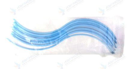
How modern equipment solves it: Sequential dilation is the primary answer. A Teflon urethral dilator set (6Fr–16Fr, 70cm) allows stepwise enlargement of the lumen with a low-friction PTFE surface that reduces mucosal trauma during passage — far gentler than forcing a scope through a resistant orifice.
Ureteral balloon dilation catheters provide an alternative for transluminal dilation of ureteral strictures, with radiopaque markers indicating balloon ends for fluoroscopic placement.
Hydrophilic and standard 0.035″ guidewires establish a safe track before any larger instrument is committed, and a smaller-caliber semi-rigid uretero-renoscope — a 6/7Fr, 430mm uretero-renoscope, for instance — can often negotiate a ureter that will not accept a larger scope at all.
2. Stone Retropulsion and Migration
The challenge: Stone migration is among the most frequently reported URS complications. During fragmentation, energy delivered to the stone can push it proximally back into the renal collecting system, converting a straightforward distal ureteric case into a retrograde intrarenal procedure — or forcing a staged second operation. The problem is worst with hard stones and aggressive energy settings.
How modern equipment solves it: Anti-retropulsion devices are the direct fix. A stone cone or nitinol retrieval coil is deployed proximal to the stone before fragmentation begins, physically blocking fragments from migrating upward and simultaneously helping sweep fragments distally on withdrawal.
On the energy side, modality choice matters: pneumatic lithotripsy is highly effective at fragmenting dense stones but carries a higher retropulsion risk than laser, which is why many surgeons use a hybrid approach — pneumatic energy for initial disintegration of a dense impacted calculus, laser for controlled finishing with minimal migration.
A modern intracorporeal pneumatic lithotripter with selectable single, multiple, and pulse modes gives the surgeon far more control over energy delivery than a fixed-output device, which is precisely how retropulsion is managed in practice.
3. Impacted Stones and Mucosal Edema
The challenge: Impacted ureteric stones, particularly in the lower ureter, are associated with prolonged operative time. Chronic impaction produces mucosal edema and inflammatory narrowing around the calculus, so the surgeon faces a stone that is both hard to reach and hard to see, embedded in tissue that bleeds and swells on contact.
How modern equipment solves it: A semi-rigid ureteroscope with a straight, adequately sized working channel allows a lithotripsy probe to be delivered directly onto the stone under direct vision without sacrificing irrigation flow. Pneumatic lithotripsy remains the workhorse for dense impacted stones because its mechanical energy is unaffected by stone composition or colour — unlike laser, whose efficiency varies with stone characteristics. Grasping forceps and biopsy forceps allow the surgeon to clear edematous tissue and assess the mucosa around the impaction site before and after fragmentation.
4. Poor Visibility and Irrigation Control
The challenge: Bleeding from an inflamed ureteric wall, stone dust clouding the field, and irrigation that drops off the moment an instrument occupies the working channel are a constant tax on operative efficiency. Poor visualization is not just an inconvenience; it directly raises the risk of ureteral wall injury and missed residual fragments.
How modern equipment solves it: A properly specified irrigation system with dedicated tubing maintains consistent flow and pressure rather than relying on gravity alone. Ureteral access sheaths in 10/12Fr or 12/14Fr sizes create a continuous outflow channel, which clears debris and blood while keeping the working field legible. On the optical side, a quality light cable paired with a camera head and monitor determines how much of the scope’s resolution actually reaches the surgeon — a sharp telescope behind a degraded light cable delivers a degraded image, which is one of the more common and most overlooked causes of poor intraoperative visibility.
5. Tortuous Anatomy and Limited Maneuverability
The challenge: Kinked, angulated, or tortuous ureters — whether from spinal deformity, prior surgery, or normal anatomical variation — reduce ureteroscopic maneuverability and increase resistance to advancement. Documented cases describe acute ureteral angulation requiring gentle torque control, minimal advancement force, and repeated realignment to avoid tearing the ureter. Lower pole renal stones present the same problem from a different angle: the access route itself fights the instrument.
How modern equipment solves it: This is where flexible ureteroscopy earns its place. A flexible ureteroscope with active deflection reaches calyces a semi-rigid scope cannot, and disposable flexible scopes remove the risk of a damaged reusable scope taking the case — and the department’s only flexible scope — out of service. A zebra-type hydrophilic nitinol guidewire offers strong visual feedback and superior handling through tortuous segments, and a ureteral access sheath, once placed, straightens the ureteric course and permits repeated scope passes without re-navigating the anatomy each time.
6. Fragment Retrieval and Residual Stone Burden
The challenge: Fragmentation is only half the job. Residual fragments are a recognised cause of recurrent symptoms and reintervention, and retrieving small, irregular, or awkwardly positioned pieces from a calyx is often the longest phase of the procedure.
How modern equipment solves it: A tipless nitinol stone basket in the 1.5–2.2Fr range is the primary retrieval tool — the tipless design allows engagement of fragments in a calyx without the basket tip traumatising the mucosa, and nitinol’s shape memory gives consistent opening behaviour across repeated deployments. Grasping forceps, both standard and long, handle larger fragments that a basket would struggle to secure. Having both available matters: a department with only baskets will spend longer on large fragments, and one with only forceps will struggle with small calyceal pieces.
7. Ureteral Wall Injury
The challenge: Ureteral wall injury sits alongside stent discomfort and stone migration as one of the three most frequently reported URS complications. It ranges from superficial mucosal abrasion to full-thickness perforation and, in the worst cases, avulsion. Most injuries trace back to force applied against resistance — a sheath pushed into a ureter too narrow for it, a basket withdrawn with a fragment too large to pass.
How modern equipment solves it: Instrument design does most of the preventive work here. Tapered atraumatic sheath tips distribute force on insertion rather than concentrating it. Smooth, uniformly tapered dilators with radiopaque construction reduce trauma while remaining visible under fluoroscopy. Tipless baskets avoid the sharp leading edge that causes mucosal tears during withdrawal. And a Double J stent (4.8–6Fr) with a proper insertion set allows the ureter to be stented and rested when injury does occur or when access proves too traumatic to continue safely.
8. Scope Damage and Repair Downtime
The challenge: This is a procurement problem that presents as a clinical one. A damaged flexible ureteroscope sent for repair can take a department’s URS capability offline for weeks, and repair costs accumulate quietly into a significant annual line item. Damage typically results from over-deflection, passing an instrument through a deflected scope, or improper sterilization handling.
How modern equipment solves it: Two complementary approaches. First, a ureteral access sheath protects the scope during repeated passes, which is one of the more reliable ways to extend flexible scope lifespan. Second, single-use flexible ureteroscopes eliminate repair downtime for departments where scope availability is the binding constraint. For reusable instruments, medical-grade construction with verified compatibility across autoclaving and low-temperature sterilization cycles is what separates a set that holds its precision over years from one that degrades within months.
9. Infection Risk and Intrarenal Pressure
The challenge: Urosepsis sits at the severe end of the URS complication spectrum. Elevated intrarenal pressure during irrigation is a recognised contributor, driving bacteria and endotoxins into the renal circulation — a particular risk in infected stone cases.
How modern equipment solves it: A ureteral access sheath is the key instrument here, providing continuous outflow that keeps intrarenal pressure low throughout the procedure while simultaneously improving visualization and easing fragment removal. Beyond the sheath, sterilization-compatible instrument construction and appropriate single-use components for the highest-risk elements of the set underpin infection control across a high-volume list.
Challenge-to-Instrument Reference Table
| Intraoperative Challenge | Primary Instrument Solution | Supporting Components |
|---|---|---|
| Narrow ureter / failed access | Teflon dilator set (6Fr–16Fr) | Hydrophilic guidewire, balloon dilator, small-caliber ureteroscope |
| Stone retropulsion | Stone cone / anti-retropulsion coil | Pneumatic lithotripter with pulse-mode control |
| Impacted stone with edema | Pneumatic lithotripter | Semi-rigid scope, grasping forceps |
| Poor visibility | Ureteral access sheath (10/12Fr, 12/14Fr) | Irrigation system, light cable, camera head |
| Tortuous anatomy | Flexible ureteroscope | Zebra nitinol guidewire, access sheath |
| Residual fragments | Tipless nitinol basket (1.5–2.2Fr) | Grasping forceps, standard and long |
| Ureteral wall injury | Atraumatic tapered sheaths and dilators | Double J stent (4.8–6Fr), stent insertion set |
| Scope damage / downtime | Ureteral access sheath, single-use flexible scope | Sterilization-compatible reusable instruments |
| Infection / high intrarenal pressure | Ureteral access sheath | Autoclavable instrument set, single-use components |
Building a Ureteroscopy Set That Handles Difficult Cases
Most of the challenges above are foreseeable. A department that stocks only for the straightforward case will meet the difficult one unprepared, and the cost shows up as prolonged operative time, staged reintervention, or an avoidable complication. A few procurement principles follow from that:
- Buy the set, not the pieces. Integrated ureteroscopy sets offer verified compatibility between components, shorter setup time, and a streamlined workflow — advantages that disappear when instruments are sourced individually from multiple vendors.
- Stock both semi-rigid and flexible capability. Semi-rigid handles the distal and mid ureter efficiently; flexible reaches the upper tract and lower pole. Departments with only one are forced into compromises on a predictable share of cases.
- Hold a range of sheath and dilator sizes. Difficult access is common enough that 10/12Fr and 12/14Fr sheaths plus a full 6Fr–16Fr dilator range should be standing stock, not a special order.
- Carry both retrieval modalities. Baskets and forceps solve different retrieval problems; the combination shortens the longest phase of most cases.
- Verify certification with documentation. ISO 13485 and CE certification should be confirmed through actual certificates, not a logo on a product page — it matters for hospital procurement approval and for import clearance in export markets.
- Check sterilization and after-sales terms. Confirm autoclave and low-temperature sterilization compatibility, expected cycle life, and whether individual components can be reordered without buying a replacement set.
Hospitals and distributors comparing a ureteroscopy set price across suppliers should weigh these factors alongside the quote itself. Sourcing from a single-source urology instruments manufacturer in India that supplies the complete set — scopes, sheaths, dilators, baskets, forceps, and the lithotripsy device together — simplifies warranty coverage, replacement-part sourcing, and bulk procurement considerably compared with assembling equivalent capability from several vendors.
FAQs
What is the most common problem during ureteroscopy?
Ureteral stent discomfort, ureteral wall injury, and stone migration are the three most frequently reported complications in the published literature. Intraoperatively, difficulty accessing a narrow ureter is among the most common reasons a case is delayed, staged, or abandoned in favour of pre-stenting.
How can stone retropulsion be prevented during ureteroscopy?
Anti-retropulsion devices such as a stone cone or nitinol retrieval coil are deployed proximal to the stone before fragmentation to block migration. Energy control also matters — a lithotripter with selectable single, multiple, and pulse modes allows the surgeon to modulate output rather than applying fixed maximum energy to a mobile stone.
Why does a ureteral access sheath matter?
A ureteral access sheath does several jobs at once: it maintains a working channel for repeated scope passes, improves visualization through continuous outflow, reduces intrarenal pressure and therefore infection risk, eases fragment retrieval, and protects the scope from wear during repeated insertion.
Should a department buy a complete ureteroscopy set or source instruments individually?
Integrated sets offer verified component compatibility, reduced setup and procedure time, and a simpler warranty and replacement pathway. Sourcing individually introduces compatibility risk that typically surfaces mid-procedure, and complicates after-sales support across multiple vendors.
What should hospitals check before buying a ureteroscopy set in bulk?
Confirm ISO 13485 and CE certification with documentation, check that the supplier provides the complete component range rather than partial sets, verify sterilization compatibility and expected cycle life, and clarify bulk pricing, lead times, export documentation, and whether individual components can be reordered separately.
AHCR (Advanced Health Care Resources) manufactures complete ureteroscopy sets, pneumatic lithotripters, and the full range of urology instruments under ISO 13485:2012 and CE certification, supplying hospitals, distributors, and urology departments worldwide. View the complete ureteroscopy set or contact us for pricing and bulk procurement enquiries.






Leave a Reply